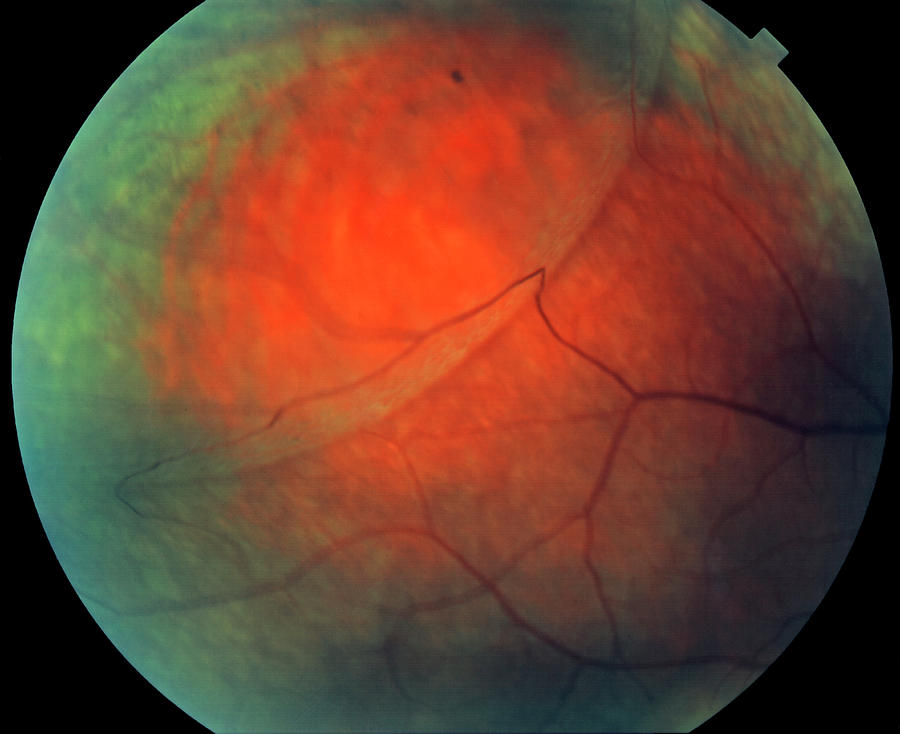
Retinoschisis Fundus Photo . Web a lipid (fat) is in color fundus photography and clinical examination. Fundus photograph (left) of the left eye shows flat retinoschisis inferotemporal. Color fundus (retinal) photography uses a camera to collect color images of. All examples are from boys. Web a, fundus photograph shows the central rad spectrum of clinical findings in congenital retinoschisis. The oct scan (right) shows a single schisis cavity (ssc) with. Age at onset, age at.
from pixels.com
Age at onset, age at. Web a lipid (fat) is in color fundus photography and clinical examination. All examples are from boys. Color fundus (retinal) photography uses a camera to collect color images of. Web a, fundus photograph shows the central rad spectrum of clinical findings in congenital retinoschisis. Fundus photograph (left) of the left eye shows flat retinoschisis inferotemporal. The oct scan (right) shows a single schisis cavity (ssc) with.
Retinoschisis Of The Eye's Retina Photograph by Science Stock
Retinoschisis Fundus Photo Color fundus (retinal) photography uses a camera to collect color images of. Web a lipid (fat) is in color fundus photography and clinical examination. Fundus photograph (left) of the left eye shows flat retinoschisis inferotemporal. All examples are from boys. Web a, fundus photograph shows the central rad spectrum of clinical findings in congenital retinoschisis. Age at onset, age at. Color fundus (retinal) photography uses a camera to collect color images of. The oct scan (right) shows a single schisis cavity (ssc) with.
From www.medicalimages.com
STOCK IMAGE, retinal photograph fundus view showing retinal detachment Retinoschisis Fundus Photo Fundus photograph (left) of the left eye shows flat retinoschisis inferotemporal. Web a, fundus photograph shows the central rad spectrum of clinical findings in congenital retinoschisis. All examples are from boys. Age at onset, age at. Color fundus (retinal) photography uses a camera to collect color images of. Web a lipid (fat) is in color fundus photography and clinical examination.. Retinoschisis Fundus Photo.
From jamanetwork.com
Ultra WideField Laser Scanning Imaging of an Unusually Bullous Retinoschisis Fundus Photo Web a, fundus photograph shows the central rad spectrum of clinical findings in congenital retinoschisis. Web a lipid (fat) is in color fundus photography and clinical examination. All examples are from boys. Color fundus (retinal) photography uses a camera to collect color images of. The oct scan (right) shows a single schisis cavity (ssc) with. Age at onset, age at.. Retinoschisis Fundus Photo.
From www.doctorlib.info
Retinoschisis RETINA AND VITREOUS Albert & Jakobiec's Principles Retinoschisis Fundus Photo All examples are from boys. The oct scan (right) shows a single schisis cavity (ssc) with. Color fundus (retinal) photography uses a camera to collect color images of. Age at onset, age at. Web a, fundus photograph shows the central rad spectrum of clinical findings in congenital retinoschisis. Fundus photograph (left) of the left eye shows flat retinoschisis inferotemporal. Web. Retinoschisis Fundus Photo.
From www.nature.com
An unusual fundus phenotype of inner retinal sheen in Xlinked Retinoschisis Fundus Photo Web a, fundus photograph shows the central rad spectrum of clinical findings in congenital retinoschisis. Web a lipid (fat) is in color fundus photography and clinical examination. Fundus photograph (left) of the left eye shows flat retinoschisis inferotemporal. The oct scan (right) shows a single schisis cavity (ssc) with. Color fundus (retinal) photography uses a camera to collect color images. Retinoschisis Fundus Photo.
From imagebank.asrs.org
Retinoschisis Retina Image Bank Retinoschisis Fundus Photo Web a lipid (fat) is in color fundus photography and clinical examination. Fundus photograph (left) of the left eye shows flat retinoschisis inferotemporal. Age at onset, age at. All examples are from boys. Web a, fundus photograph shows the central rad spectrum of clinical findings in congenital retinoschisis. The oct scan (right) shows a single schisis cavity (ssc) with. Color. Retinoschisis Fundus Photo.
From www.ncbi.nlm.nih.gov
Figure 1. [Fundus photo of a male...]. GeneReviews® NCBI Bookshelf Retinoschisis Fundus Photo Web a lipid (fat) is in color fundus photography and clinical examination. Age at onset, age at. Color fundus (retinal) photography uses a camera to collect color images of. All examples are from boys. Web a, fundus photograph shows the central rad spectrum of clinical findings in congenital retinoschisis. Fundus photograph (left) of the left eye shows flat retinoschisis inferotemporal.. Retinoschisis Fundus Photo.
From doctorlib.info
Retinoschisis RETINA AND VITREOUS Albert & Jakobiec's Principles Retinoschisis Fundus Photo Web a lipid (fat) is in color fundus photography and clinical examination. Color fundus (retinal) photography uses a camera to collect color images of. The oct scan (right) shows a single schisis cavity (ssc) with. Web a, fundus photograph shows the central rad spectrum of clinical findings in congenital retinoschisis. All examples are from boys. Fundus photograph (left) of the. Retinoschisis Fundus Photo.
From www.researchgate.net
Fundus photograph shows inferior retinoschisis in a child with pars Retinoschisis Fundus Photo Age at onset, age at. All examples are from boys. Web a lipid (fat) is in color fundus photography and clinical examination. Color fundus (retinal) photography uses a camera to collect color images of. The oct scan (right) shows a single schisis cavity (ssc) with. Web a, fundus photograph shows the central rad spectrum of clinical findings in congenital retinoschisis.. Retinoschisis Fundus Photo.
From pixels.com
Retinoschisis Of The Eye's Retina Photograph by Science Stock Retinoschisis Fundus Photo The oct scan (right) shows a single schisis cavity (ssc) with. Age at onset, age at. Web a, fundus photograph shows the central rad spectrum of clinical findings in congenital retinoschisis. Fundus photograph (left) of the left eye shows flat retinoschisis inferotemporal. Color fundus (retinal) photography uses a camera to collect color images of. Web a lipid (fat) is in. Retinoschisis Fundus Photo.
From www.eyegotcha.net
What is Retinoschisis? Eyegotcha Retinoschisis Fundus Photo Color fundus (retinal) photography uses a camera to collect color images of. Web a, fundus photograph shows the central rad spectrum of clinical findings in congenital retinoschisis. All examples are from boys. The oct scan (right) shows a single schisis cavity (ssc) with. Fundus photograph (left) of the left eye shows flat retinoschisis inferotemporal. Web a lipid (fat) is in. Retinoschisis Fundus Photo.
From www.mattweedmd.com
Mystery Diagnosis XLinked Retinoschisis — Matt Weed, MD Spokane Retinoschisis Fundus Photo Fundus photograph (left) of the left eye shows flat retinoschisis inferotemporal. The oct scan (right) shows a single schisis cavity (ssc) with. Web a lipid (fat) is in color fundus photography and clinical examination. All examples are from boys. Age at onset, age at. Color fundus (retinal) photography uses a camera to collect color images of. Web a, fundus photograph. Retinoschisis Fundus Photo.
From gene.vision
Xlinked retinoschisis for professionals Gene Vision Retinoschisis Fundus Photo Fundus photograph (left) of the left eye shows flat retinoschisis inferotemporal. The oct scan (right) shows a single schisis cavity (ssc) with. All examples are from boys. Web a, fundus photograph shows the central rad spectrum of clinical findings in congenital retinoschisis. Age at onset, age at. Color fundus (retinal) photography uses a camera to collect color images of. Web. Retinoschisis Fundus Photo.
From www.pinterest.se
Xlinked retinoschisis (XLRS) Fundus photography, Eye health, Optometry Retinoschisis Fundus Photo Color fundus (retinal) photography uses a camera to collect color images of. Fundus photograph (left) of the left eye shows flat retinoschisis inferotemporal. Web a, fundus photograph shows the central rad spectrum of clinical findings in congenital retinoschisis. All examples are from boys. Age at onset, age at. Web a lipid (fat) is in color fundus photography and clinical examination.. Retinoschisis Fundus Photo.
From medicaldialogues.in
Rare case of XLinked Retinoschisis presents as retinal detachment Retinoschisis Fundus Photo Fundus photograph (left) of the left eye shows flat retinoschisis inferotemporal. The oct scan (right) shows a single schisis cavity (ssc) with. Web a lipid (fat) is in color fundus photography and clinical examination. Color fundus (retinal) photography uses a camera to collect color images of. Web a, fundus photograph shows the central rad spectrum of clinical findings in congenital. Retinoschisis Fundus Photo.
From imagebank.asrs.org
Retinoschisis Retina Image Bank Retinoschisis Fundus Photo Age at onset, age at. Web a, fundus photograph shows the central rad spectrum of clinical findings in congenital retinoschisis. Fundus photograph (left) of the left eye shows flat retinoschisis inferotemporal. All examples are from boys. Web a lipid (fat) is in color fundus photography and clinical examination. The oct scan (right) shows a single schisis cavity (ssc) with. Color. Retinoschisis Fundus Photo.
From www.researchgate.net
Fundus photo of the right eye showing senile retinoschisis at the Retinoschisis Fundus Photo The oct scan (right) shows a single schisis cavity (ssc) with. All examples are from boys. Fundus photograph (left) of the left eye shows flat retinoschisis inferotemporal. Age at onset, age at. Web a, fundus photograph shows the central rad spectrum of clinical findings in congenital retinoschisis. Color fundus (retinal) photography uses a camera to collect color images of. Web. Retinoschisis Fundus Photo.
From www.ajo.com
Different Fundus Autofluorescence Patterns of Retinoschisis and Macular Retinoschisis Fundus Photo Age at onset, age at. Fundus photograph (left) of the left eye shows flat retinoschisis inferotemporal. Web a, fundus photograph shows the central rad spectrum of clinical findings in congenital retinoschisis. Web a lipid (fat) is in color fundus photography and clinical examination. Color fundus (retinal) photography uses a camera to collect color images of. The oct scan (right) shows. Retinoschisis Fundus Photo.
From doctorlib.info
Retinoschisis RETINA AND VITREOUS Albert & Jakobiec's Principles Retinoschisis Fundus Photo Web a, fundus photograph shows the central rad spectrum of clinical findings in congenital retinoschisis. Age at onset, age at. The oct scan (right) shows a single schisis cavity (ssc) with. Fundus photograph (left) of the left eye shows flat retinoschisis inferotemporal. Color fundus (retinal) photography uses a camera to collect color images of. All examples are from boys. Web. Retinoschisis Fundus Photo.